
Understanding Headaches: Causes, Types, and Relief Strategies
Headaches are the most common pain disorder worldwide, affecting roughly 66% of the global population. The International Headache Society classifies headaches into several categories:Tension-type headachesCervicogenic headachesMigraine and cluster headachesSecondary headaches resulting from underlying conditions (e.g., fever, infection, sinus issues, or tumors)Cranial neuralgias, facial pain, and other headache syndromes

Tension-Type Headaches
Tension-type headaches are the most common form of headache in adults, with studies suggesting that 30–80% of U.S. adults experience them. These headaches often result from stress, poor posture, fatigue, jaw or neck pain, or dysfunctional breathing patterns.People often describe the pain as a dull ache or tight, band-like pressure around the head — particularly at the temples, forehead, or the back of the neck. The discomfort can last anywhere from 30 minutes to several days and usually affects both sides of the head, although it may occasionally occur on one side.Anatomically, this pain occurs because the neck muscles attach to the cervical spine and skull. When these muscles tighten or spasm, they can “tug” on the skull and create tension that radiates into different regions of the head. Depending on which muscles are involved, pain may be felt at the base of the skull, across the forehead, or above the eyes.Common Muscles That Contribute to Tension Headaches
Upper TrapeziusSternocleidomastoidLevator ScapulaeSuboccipitalsScalenes
These muscles, when tight or overused, can refer pain into the head and mimic classic headache symptoms.
Managing Tension Headaches
Over-the-counter medications such as Tylenol, Advil, Motrin, or Aleve can help relieve symptoms, but it’s important to consult your doctor before use. Always take the lowest effective dose, as overuse can sometimes lead to rebound headaches — a worsening cycle of pain triggered by frequent medication use.
Cervicogenic Headaches
Unlike tension headaches, cervicogenic headaches stem from dysfunction in the upper three cervical spine segments (facet or zygapophyseal joints). Nerves originating in the cervical spine provide sensory and motor information to the head and neck, and irritation of these nerves can produce referred pain that mimics a headache.
Common Identifiers of Cervicogenic Headaches:
Pain primarily on one side of the head (though both sides can be affected)Restricted neck movement or stiffnessPain triggered by neck movement or awkward posturesPain reproduced by pressure at the base of the skull or upper cervical spineAccompanying same-sided neck, shoulder, or arm pain
Treatment and Outlook
Research shows that neck-specific exercises — particularly low-intensity endurance training — and spinal mobilization can be effective both in the short and long term for managing cervicogenic headaches.It’s also important to note that not all headaches share the same cause. Some result from dehydration, lack of sleep, or underlying medical issues. Identifying the root cause is the first step toward effective relief — and many cases can be managed without heavy reliance on medication.In some instances, a local anesthetic injection may be required for pain relief, but this is typically reserved for more severe or persistent cases. Physical therapists can play a crucial role in addressing both tension-type and cervicogenic headaches through posture correction, strengthening, and manual therapy. When in doubt, consult a neurologist or primary care physician to help determine the underlying source of your headache.Treatment Strategies:
1. Posture Correction & Mobility Exercises
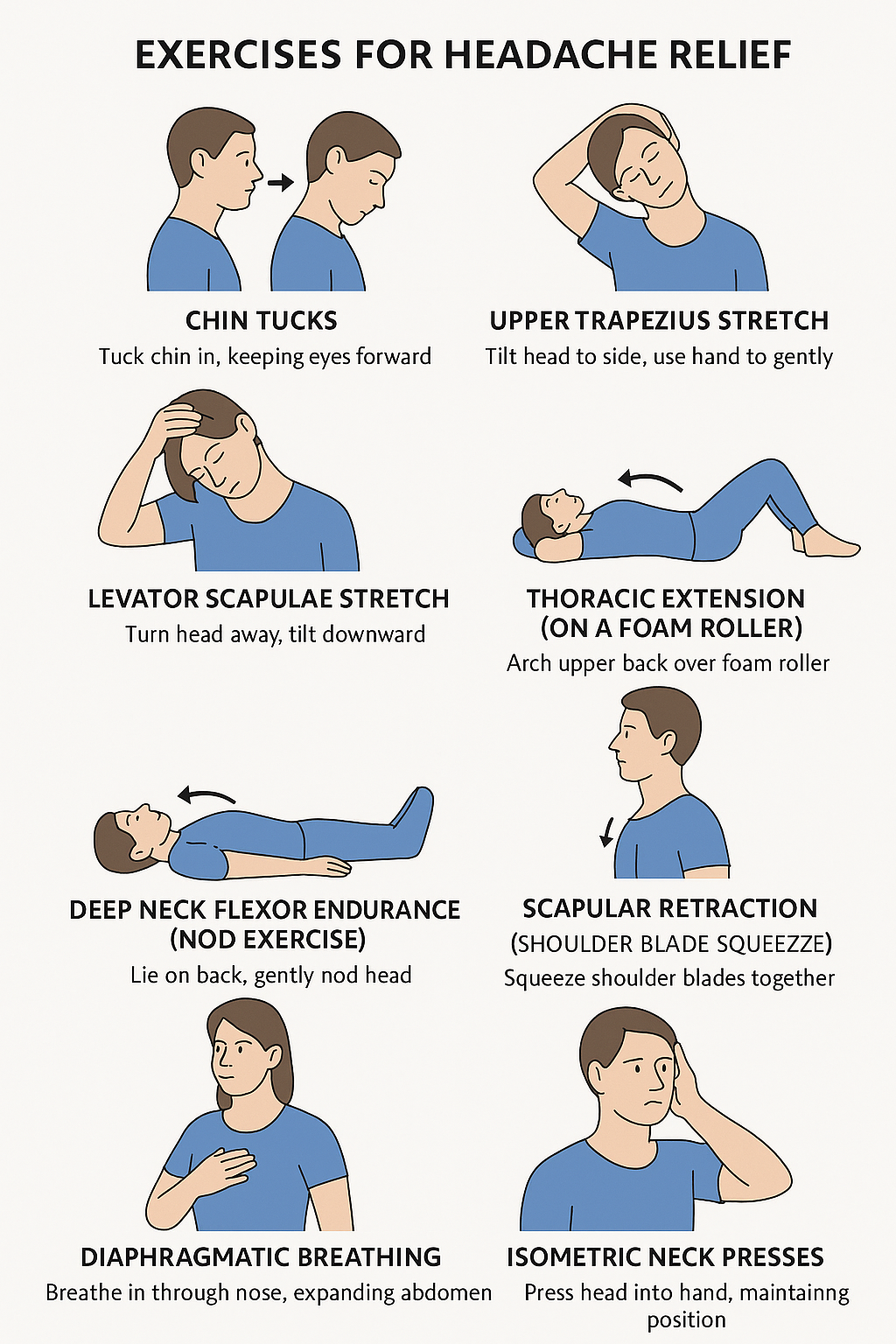
These target stiffness and poor alignment — common triggers for both tension-type and cervicogenic headaches.Chin Tucks
How: Sit or stand tall. Gently draw your chin straight back (as if making a double chin), keeping your eyes level.Hold: 5 secondsReps: 10–15Goal: Strengthen deep neck flexors and counteract forward-head posture.
Upper Trapezius Stretch
How: Sit tall. Gently tilt your head to one side (ear toward shoulder). Use your hand to lightly increase the stretch.Hold: 20–30 seconds each sideReps: 2–3 per sideGoal: Reduce tightness in the upper trapezius muscles.
Levator Scapulae Stretch
How: Sit tall. Turn your head 45° to one side, then look down toward your armpit. Use your hand to apply gentle pressure on the back of your head.Hold: 20–30 seconds each sideReps: 2–3 per sideGoal: Decrease neck base and shoulder tension.
Thoracic Extension (on a Foam Roller)
How: Lie with a foam roller under your upper back, knees bent. Support your head and gently arch backward over the roller.Hold: 2–3 secondsReps: 10–12Goal: Improve upper-back mobility and reduce strain on the neck.
2. Neck Strengthening & Endurance Training
These build stability in deep neck muscles that often weaken due to poor posture or prolonged screen time.Deep Neck Flexor Endurance (Nod Exercise)
How: Lie on your back with knees bent. Gently nod your head (“yes” motion) without lifting it off the floor.Hold: 5–10 secondsReps: 10–12Goal: Strengthen deep cervical flexors — crucial for posture and neck support.
Scapular Retraction (Shoulder Blade Squeeze)
How: Sit or stand tall. Gently squeeze your shoulder blades together and down (avoid shrugging).Hold: 5 secondsReps: 10–15Goal: Strengthen mid-back muscles that support the neck.
Isometric Neck Presses
How: Press your head gently into your hand (forward, backward, side to side) without actual movement.Hold: 5 seconds each directionReps: 5–10 each wayGoal: Build neck stability and reduce muscular fatigue.
3. Breathing & Relaxation Techniques
Dysfunctional breathing and stress can amplify headache frequency and intensity.Diaphragmatic Breathing
How: Sit or lie comfortably. Place one hand on your belly and one on your chest. Inhale deeply through your nose so your belly rises, then exhale slowly through your mouth.Duration: 2–5 minutesGoal: Reduce muscle tension and activate relaxation responses.
4. Daily Habit Fixes
Simple lifestyle adjustments can make these exercises even more effective:Take movement breaks every 30–45 minutes during desk work.Keep your monitor at eye level to reduce neck strain.Stay hydrated and get adequate sleep.Consider gentle yoga or Pilates to maintain flexibility and posture control.
Understanding Hamstring Injuries in Sports: How Common Are They—and What You Can Do
Hamstring injuries are one of the most common complaints among athletes—especially in sports involving sprinting, kicking, or sudden stops and starts. If you’ve ever pulled up with a tight back thigh or heard the dreaded “pop,” you know just how disruptive these injuries can be. In this post, we’ll look at what the research says about how often hamstring injuries happen, who’s at risk, and what physical therapy can do to prevent and recover from them.

Hamstring injuries are one of the most common complaints among athletes—especially in sports involving sprinting, kicking, or sudden stops and starts. If you’ve ever pulled up with a tight back thigh or heard the dreaded “pop,” you know just how disruptive these injuries can be. In this post, we’ll look at what the research says about how often hamstring injuries happen, who’s at risk, and what physical therapy can do to prevent and recover from them.What Are Hamstring Injuries?
The hamstrings are a group of muscles and tendons at the back of the thigh. Injuries range from mild strains (small tears) to more serious ruptures or tendon injuries. Some are acute (sudden) while others develop over time or recur.How Common Are They?
Here are some key statistics from recent studies:Incidence in field‑based team sportsAbout 0.81 hamstring injuries per 1,000 exposure hours (training + matches) in sports like soccer, rugby, field hockey, Gaelic football, Australian football. PubMedProportion of all sports injuriesHamstring injuries make up ~10% of all injuries in those same sports. PubMedPrevalence over a season (~9 months)Around 13% of athletes will suffer a hamstring injury during a typical 9‑month season in field‑based team sports. PubMedChange over time (professional male soccer) In one 21‑season study in European male professional soccer: hamstring injuries went from accounting for ~12% of all injuries in early seasons to 24% in more recent seasons. The number of days players miss from these injuries also doubled. PubMed+1
Risk Factors: Who’s More Likely to Get Them?
During matches vs training: Hamstring injuries are much more likely during matches than training. In one review, match injury incidence was ~9.4‑fold higher than training. PubMedAge: Risk increases with age; older athletes have higher rates of hamstring injury. PubMedSurface type: Injuries are more common on natural grass vs artificial turf. PubMedPrevious injury: Athletes who have had hamstring injuries before are at greater risk of recurrence. JOSPT+2JOSPT+2
Why Are Hamstring Injuries Getting More Common?
The studies suggest a few trends:Increasing match intensity and schedules (more games, less rest). PubMed+2JOSPT+2Greater demands during training sessions. PubMed+1Possibly more awareness/reporting and better diagnostic tools, which may increase recorded incidence.In professional soccer, the “burden” (how many days missed, how long players are out) of hamstring injuries has increased over time. PubMed+1
How Physical Therapy Helps: Prevention & Recovery
Here’s how a physical therapist can help reduce risk and get you back in the game.Screening & AssessmentAssess strength, flexibility, muscle imbalances.Examine movement patterns, running or sprint mechanics.
Targeted Strength TrainingEccentric training (lengthening under load) is especially effective for hamstrings.Balance between hamstrings and quadriceps/groin muscles.
Flexibility & MobilityRegular hamstring stretches and dynamic warm‑ups.Also hip flexor and glute work to ensure pelvis positions are optimal.
Load ManagementGradually increasing training intensity.Ensuring adequate rest between high‑intensity efforts.
Neuromuscular Control & TechniqueDrills for proper technique in sprinting, cutting, stopping.Core stability, pelvic control, coordination.
Return‑to‑Sport PlanningGradual return with progressive demands.Monitoring for signs of fatigue or tightness.Ensuring full recovery before resuming sprinting or cutting loads.
Takeaway
Hamstring injuries are frequent, costly, and appear to be increasing in both number and severity—especially in sports with high demands like sprinting, football/soccer, rugby, etc. But they’re not inevitable. With careful prevention, good rehab, and smart training loads, many athletes can reduce their risk and recover fully if injury does happen.Let us know if you have any questions. We are happy to help!Shoulder Instability: Do you have Multidirectional Instability?
Do you commonly feel like your shoulder is unstable or “pops” in and out of place? If you do then you may have something called Multidirectional Instability (MDI).
Do you commonly feel like your shoulder is unstable or “pops” in and out of place? If you do then you may have something called Multidirectional Instability (MDI).
MDI is defined as laxity in the shoulder joint in multiple directions.MDI is either caused by a traumatic or atraumatic mechanism. That means the individual either had: 1. A traumatic incident like a fall, which leads to dislocation of the shoulder joint. This dislocation ultimately leads to residual laxity in the shoulder OR 2. They are a generally hypermobile individual, perform repeated overhead movements (such as with volleyball, baseball, weight lifting, etc), and have a history of subluxationsWhat is the Difference between a Subluxation and Dislocation?A Dislocation is considered a displacement of the bone from its natural position in the joint. The humeral head completely comes out of place and may require medical intervention to return the bone back into place.A Subluxation is a partial dislocation of the joint. Typically the shoulder will feel like it pops out of place but then will quickly pop back into place.
Common MDI Presentation:
<35 years of ageTypically uses words like “double jointed” or “always been flexible” to describe themselvesMultiple episodes of subluxationPain or feeling of instability typically at the end range of motionHistory of participating in overhead sports (MDI may be related to repeated microtrauma)
The good news is nonsurgical, conservative care is frequently recommended for MDI. The bad news is that these individuals will need to be diligent with a strengthening program to reduce the risk of subluxations in the future. I have seen many clients with MDI and the key is to strengthen the rotator cuff, scapulo-thoracic complex, core and hips. Addressing Range of Motion should NOT be the focus for these individuals. You do not want to create further instability by stretching the joint. Trigger point release is okay however to alleviate pain caused by overuse of the muscles associated with instability. The exercises below are a great way to start addressing MDI. Let me know if you have further questions at rachel@jsportsrehab.com!
Band Resisted Shoulder External Rotation (Bilateral):
Band Resisted Internal Rotation:
Band Resisted External Rotation:
Wall Walks:
Supine Arm Bar:
Anterior Hip Pain: Do you have Femoroacetabular Impingement (FAI)?
A recent case: A 19 year old female soccer player presented to therapy with pain into the front of her right hip and groin. She is a right footed soccer player and reported a pinch-like pain into the front of her hip as her leg traveled across her body when shooting. She also felt pain when hugging her knee to her chest and an aching pain into the hip after sitting for a few hours. She kept playing through the pain and reported the pain just seemed to get worse and worse. Clinical testing suggested she had hip impingement but Imaging from a physician officially diagnosed her with a pincer type of impingement. What is FAI?
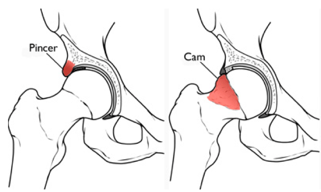
An Overgrowth of either the femoral head or the acetabulum (hip socket). It is defined as motion or position related pain in the hip or groin with possible clicking, catching, locking, stiffness, giving way, and decreased range of motion.2 Types of FAI:1. CAM: Overgrowth of Bone at the Femoral Head2. Pincer: Overgrowth of Bone at the AcetabulumCAM Impingement is more commonly seen in males in their 20s vs Pincer impingement is more commonly seen in females in their 30s and 40s.Both types of impingement can feel like a pinch. That is because there is an overgrowth of bone in the hip joint. The head of the femur and the hip socket basically bump into each other during any motions where you bring your hip up to your chest. This “bumping” or frictioning of bone on bone is not only painful, but can lead to damaging the labrum over time. The labrum is what cushions the femoral head within the socket. Damage to the labrum can potentially lead to longer recovery times or even surgery. 
Symptoms:
Pain or aching (usually located at the inner hip, or groin area), usually after walking, or prolonged sittingA locking, clicking or catching sensation within the jointPain bringing your hip up towards your chest and and across the body towards the opposite shoulder
How to Diagnose:
Clinical Testing: FADIR and FABERRange of Motion: Limitations in hip Internal rotation and flexionImaging: X rays, Magnetic Resonance Athrography (MRA), or CT scan
Treatment:
Conservative Treatment: Physical Therapy/Activity Modification: Avoid activities that cause pinching/pain– Activity Modification: Avoid activities that cause pinching/pain– Rehab Focus: Address hip strength, hip stability, neuromuscular control, range of motion, and movement mechanicsSurgical Intervention: Hip arthroscopy or open surgery: recovery time 4-6 months
Sports you are more likely to see impingement in:
Soccer, martial arts, ballet, cycling, rowing, golf, tennis, football, hockey, baseball, lacrosse, rugby, and deep squatting activities such as powerlifting.
Interesting Fact:
CAM impingement is common in male soccer players, with 60-75% of adult male soccer players found to have this type of impingement. This number is high especially when considering the prevalence of CAM in the general person is only roughly 17-20%.
Exercise Ideas:
Hip Flexor Release: https://www.youtube.com/watch?v=f13QETYZQMgQuadriceps Stretch:Couch Stretch: https://www.youtube.com/watch?v=tABeNRBDf30Bridge: https://www.youtube.com/watch?v=gYVVdmARLuUClamshell: https://www.youtube.com/watch?v=2OhR279AWNYSide Plank: https://www.youtube.com/watch?v=TPp92f8HlG0Single Leg Bird Dog: https://www.youtube.com/watch?v=GlutdS3NdXQThoracic Mobility: Important for Every Sport and Movement in Life!
Do you have low back pain, shoulder pain, or neck pain? Do you want to improve your golf swing, overhead serve in volleyball, hitting power in baseball and softball, or pitching velocity?
Do you have low back pain, shoulder pain, or neck pain? Do you want to improve your golf swing, overhead serve in volleyball, hitting power in baseball and softball, or pitching velocity?

As a physical therapist, the thoracic spine is one of first areas I treat. Whenever you lift your arm overhead, throw a ball, swing a golf club, or even look up at a book on a high shelf, your thoracic spine is moving.Try this: relax your upper back and shoulders and get really slouchy in your chair. Now try looking up at the ceiling. Then try lifting your arm overhead… Hard right? Now sit up nice and tall in your chair and perform the same two movements. Should be much easier, more comfortable, and you should be able to get more range of motion in your neck and shoulder. I know this is a very extreme example but imagine how much even a little bit of thoracic spine stiffness can affect how you get your arms overhead to spike a ball in volleyball or throw a baseball.Common problems associated with poor thoracic mobility include shoulder impingement/pain, low back pain, and neck pain. There are other factors that can contribute to these same issues but, in this article, we are going to focus on the thoracic spine as the root of the problem. When the thoracic spine doesn’t move, your low back starts to pick up the slack. Instead of getting normal extension through your thoracic spine you end up hyperextending through your lumbar spine. This repetitive lumbar hyperextension leads to pinching posteriorly of the bones in your low back, thus resulting in pain. The same issues can happen at your neck.
When you lift your arm overhead and you cannot extend in the thoracic spine, what will typically happen is your humerus will jam up against the acromion (See picture below). Having this bone to bone contact overtime can lead to pain. This same issue can also lead to rotator cuff muscle microtears because the rotator cuff muscle sits right underneath the acromion, and the friction of the constant bone to bone contact can lead to fraying of the muscle underneath.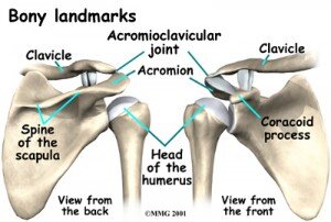
From a performance perspective in overhead athletes or overhead lifting (push press, push jerk, overhead squat), more force can be generated and more weight can be lifted when you have the range of motion to complete the movement. To properly engage your shoulder blade muscles and have your shoulder blade sit properly on the ribcage, you need full range of motion of the shoulder joint and thoracic spine. If not, like we mentioned before, your shoulder blades will sit forward on your ribcage (like with bad slouching posture) and your shoulder blade muscles won’t be able to engage properly. The better your shoulder blade muscle strength and stability are, the more power you can generate and the more weight you can lift overhead.Imagine swinging a baseball bat if you can’t rotate your thoracic spine. With this limitation the force you can produce when you hit the ball will be considerably less as compared to being able to rotate your spine fully.Here are some exercises you can do on your own to address this area of the body:7 Common Reasons Why You Have Shoulder Pain
Shoulder pain is one of my favorite issues to treat. For me, shoulder pain is easy to treat. Why? Because there are a lot of commonalities among individuals that come to me with shoulder pain, no matter what type of shoulder pain it is. No, not everyone with shoulder pain will have the same exact 3 or 4 problems; which is why an extensive examination is necessary before I start treating someone. However, I still find that most of my shoulder pain patients have a few things in common. Listed below are the top 7 dysfunctions I see in my shoulder patients:1. Restricted pectoralis minor/major flexibilityYou will notice difficulty reaching behind your back
2. Restricted latissimus dorsi flexibilityYou will notice difficulty raising your arm to your ear
3. Trigger points (muscle knots) that refer pain into the shoulderIf you apply deep pressure to certain muscles, you will feel tender points that refer pain elsewhere (especially common with the rotator cuff muscles that sit on top of your shoulder blade)
4. Forward head and shoulder postureMost people have sitting jobs and tend to sit with slouchy posture
5. Overactivation of the upper trapezius musclesYou will notice you constantly shrug your shoulders up when lifting your arm overhead
6. Weak scapular retractors, core, and rotator cuff muscles
7. Poor motor control
You have the range of motion but your body doesn’t engage the right muscles at the right time appropriately to perform a specific movement
Next I want to talk about some common diagnoses I hear when a patient walks into my facility.Pain in the biceps or the front of the shoulder: A lot of times people come in with pain in the front of the shoulder and have been told they have a biceps tendonitis/tendonapathy. If you hear this diagnosis it means your biceps tendon has taken on extra work because your rotator cuff muscles, your scapular stabilizers and your core are slacking on the job. Other common issues found with this diagnosis include: restricted thoracic mobility, and slouchy forward head and shoulder posture, tight pectoralis minor/major muscles, and potentially tight latissimus dorsi muscles.
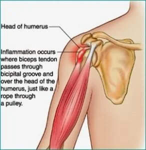
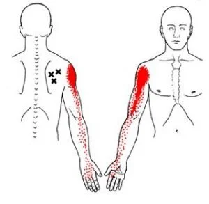
Pain that travels from the top of your shoulder down your arm an inch or two? This sensation is commonly due to what we call active trigger points (inflamed muscle knots). Your rotator cuff muscles, which sit on top of the shoulder blade, can develop trigger points that can refer pain into the shoulder and, at times, down the arm. Performing soft tissue massage using the lacrosse ball on tender or tight spots will help calm down the muscle and reduce the feeling of pain traveling into your shoulder.
Pain at the top of the shoulder joint and pain with raising your arm to end range overhead: This type of pain is typically given the diagnosis of shoulder impingement/ bursitis. This occurs because the humerus (arm bone) basically jams into the top of the shoulder joint whenever you raise your shoulder overhead. These people typically have bad forward head and shoulder posture. This diagnosis is either due to excessive shoulder mobility, or it is due to tight shoulder musculature (pectoralis minor/major, lats, rotator cuff muscles), overuse of the upper trapezius muscles, restricted thoracic mobility, or the inability to use your back, shoulder blade and core muscles efficiently. The latter option I tend to see more often.

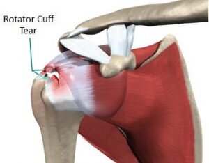
Shoulder pain that is associated with weakness and, at times, the inability to lift your arm overhead: This injury can result from a traumatic fall or gradual wear and tear of the muscle with overhead activity. Common issues I see with this diagnosis include: forward head and shoulder posture, restricted thoracic mobility, tight shoulder musculature (pectoralis minor/major, latts, Rotator Cuff muscles), overuse of the upper trapezius muscles, and you are not using your back, shoulder blade and core muscles efficiently.
Notice a trend with some of the different diagnoses?.. It may seem like I am repeating myself a lot but most shoulder diagnoses stem from the same problems (excluding traumatic shoulder injuries of course). If you have shoulder pain and are reading this article then attacking the 7 problems I have listed above is a good place to start.Patellar Tendinopathy (Jumpers Knee):
Have you ever had pain in the front of the knee with jumping, cutting, running, or kicking? I personally have and it stinks. I played soccer growing up so the feeling of a sharp jolt to the front of the knee is not something foreign to me. To those of you experiencing the same sharp pain to the front of the knee, you will know that it feels as if you are running or jumping on eggshells.
What Is It and How Do You Treat It?
Have you ever had pain in the front of the knee with jumping, cutting, running, or kicking? I personally have and it stinks. I played soccer growing up so the feeling of a sharp jolt to the front of the knee is not something foreign to me. To those of you experiencing the same sharp pain to the front of the knee, you will know that it feels as if you are running or jumping on eggshells. You want to be explosive but you feel like if you bend your knees too much to really generate power or to absorb a landing, the sharp jolt is just waiting for you not far behind. I experienced this pain as a soccer player but it affects many other sports such as basketball, volleyball, tennis, track and field, football and the recreational runner. Athletes between the ages of 15 to 30 are primarily affected, with men more commonly affected than women. By definition, patellar tendinopathy (Jumper’s knee) is an overuse injury thought to be caused by excessive or repetitive forces applied to the patellar tendon. Signs and Symptoms of Patellar Tendinopathy:
Anterior knee pain over the patella tendonTenderness at the inferior pole of the patellar tendonPain made worse with jumping, landing, cutting/pivoting, or running activitiesPain with stairs, squatting, and at times prolonged sittingOnset of pain is usually gradual and commonly related to an increase in sport activityThe feeling of stiffness in the patellar tendon in the morningIncreased thickness of the patellar tendon compared to the opposite side
What is the Function of the Patellar Tendon?
The patellar tendon attaches directly from the tibial tuberosity to the inferior tip of the patella (knee cap). The quadriceps muscle attaches into the superior tip of the patella, thus transmits forces through the patellar tendon based on the common attachment into the patella. Therefore, any tightness or weakness in the quadriceps pulls on the patellar tendon. The patellar tendon also functions in the storage and release of energy when you jump, run, cut or pivot. This means there is a lot of load that has to transfer through the patellar tendon so overtraining, insufficient rest, faulty movement patterns, mobility impairments, and hip, core and foot weakness can contribute to the development of patellar tendinopathy.
Common Impairments Found in Individuals with Patellar Tendinopathy:
Hip Abductor and Hip External Rotator WeaknessAltered Hip and Knee Movement Patterns upon Landing (Poor Movement Quality)Excessive or Restricted Hip and Ankle MobilityLook for reduced dorsiflexion, quadriceps or hamstring tightness, hip Internal rotation mobility, increased varus foot alignment, etc.
Quadriceps WeaknessExcessive Foot Pronation: foot collapses inward upon landing which causes knee valgus (knee collapse inward) as well as the femur to then internally rotate excessively
Rehabilitation Focus:
Eccentric Quadriceps StrengtheningQuadriceps and Hamstring FlexibilityHip Abduction and External Rotation StrengtheningMovement Competency–May require Cueing from a Rehab Professional or Qualified Strength Coach to address running, cutting, and jumping mechanicsFoot and Ankle StrengtheningAnkle Dorsiflexion Range of Motion
Exercise Ideas:
Here are some exercise ideas to get you started but it is always helpful to seek help from a medical professional.Quadriceps Flexibility:Couch Stretch: https://www.youtube.com/watch?v=tABeNRBDf30
Hip External Rotation Strengthening:Clamshell: https://www.youtube.com/watch?v=2OhR279AWNYFire Hydrant (can add band around knees to increase difficulty): https://www.youtube.com/watch?v=0Vk6BrtikL8Side Plank with Clamshell: https://www.youtube.com/watch?v=82tKGsLrADY
Hip Abduction Strengthening:Sidelying Hip Abduction: https://www.youtube.com/watch?v=Ku6QkDJqdwYSide Plank: https://www.youtube.com/watch?v=TPp92f8HlG0
Ankle Dorsiflexion:Mobilization with Movement: Long Sitting Position with Mobility Band: https://www.youtube.com/watch?v=sBCMIaIkP8cWall Stretch: https://www.youtube.com/watch?v=9QRVlaw9QWM
References:
Malliaras, P., Cook, J., Purdam, C. and Rio, E. (2015). Patellar Tendinopathy: Clinical Diagnosis, Load Management, and Advice for Challenging Case Presentations. Journal of Orthopaedic & Sports Physical Therapy, 45(11), pp.887-898.
Mendonça, Luciana D., et al. “Association of Hip and Foot Factors With Patellar Tendinopathy (Jumper’s Knee) in Athletes.” Journal of Orthopaedic & Sports Physical Therapy, vol. 48, no. 9, 2018, pp. 676–684., doi:10.2519/jospt.2018.7426.
Baseball Specific Screening
The OnBase University screening stems from the belief that there is not only one technically correct way to play baseball or softball. There are an infinite number of ways to play the game. The best baseball players in history don’t all hit or pitch the same way, but they do have some commonalities in their styles that have allowed them to be the best. The aim of a good coach or trainer is to find the one efficient style that works for that player, and that is based on what the player can physically do. If we can match an athlete’s physical ability to his/her technical skills, it will help the player play at his/her highest level possible.Coach and Player Goals: To be Efficient and PowerfulWhat makes a hitting or pitching style Efficient?
It is ReproducibleThe player has command over their pitching/hitting styleThe player is able to maximize his/her power with the least amount of effort
What makes a hitting or pitching style Powerful?
Proper Kinematic Sequencing: how energy is transferred from the ground through the body to the ball or bat
It’s up to the coaches to guide the player into what kind of hitting or pitching style the athlete uses. Our goal is to assist the coach and player by determining how the athlete’s physical abilities relate to their technical skills. Pitching and hitting requires involvement from the whole body, from the feet all the way through to the neck, shoulders, elbow and hand. If there is a breakdown anywhere in this chain, it could lead to poor power, poor contact on the ball for hitters, inconsistency, and pain or injury. How is your body limiting your performance and where exactly are the breakdowns? That’s what we want to find out.How do we determine where the breakdowns are?Through Movement Screening: Functional Movement Screening (FMS), Y balance testing, OnBase University pitching and hitting screening, VALD forceplate testing, dynamometer and joint by joint assessment.We will provide these findings to you, your pitching or hitting coach, and your fitness coach to develop a team approach in order to help you become a better athlete.Let us help you and your coaches. Call us at 407-718-0437 or email us at rachel@jsportsrehab.com..Reference: http://www.onbaseu.com